Home » Infographic » Quelles sont les causes principales de l’apparition de la vallée des larmes ?
The “tear trough” gives the eyes a tired, sad, or aged appearance. This anatomical area, also called the nasolacrimal groove, is particularly delicate because it lies at the junction of multiple muscular, fatty, and bony structures. Its progressive alteration is a common concern, especially in Geneva, where a demanding clientele seeks targeted, effective, and harmonious facial treatments. Understanding the precise causes of the tear trough’s appearance is essential to offer suitable treatments and achieve natural, long-lasting results.
With age, the gradual loss of the deep fat compartments of the face is one of the main causes of the appearance of the tear trough. More specifically, the malar fat — located in the cheek area — decreases and migrates, leading to a loss of suborbital support. This fat loss is often accompanied by ptosis (sagging) of the tissues, accentuating the hollow between the orbit and the cheek. The loss of volume is not limited to superficial tissues : the deeper areas are also affected, altering the overall architecture of the face.
Bone aging also contributes to the formation of the tear trough. 3D imaging studies have shown that the malar bone (the cheekbone) gradually resorbs with age, particularly in its medial portion. This resorption reduces the structural support of the soft tissues, causing them to sag downward and accentuating the under-eye hollows. This phenomenon, often subtle but progressive, contributes over time to the perception of a tired look.
The infraorbital region is a particularly complex anatomical area, where several tissue layers with distinct functions and characteristics overlap : the epidermis, dermis, orbicularis muscle, superficial and deep fat pads, supporting ligaments, as well as the zygomatic bone and orbital rim. This multilayered, thin, and dense organization is designed to allow both high mobility and expressiveness of the eyes.
Over time, the different tissue layers lose their functional cohesion. The network of fibrous septa anchoring the superficial fat to the deep fat becomes looser, causing migration and loss of fat volumes. At the same time, the skin thins, making the underlying anatomical discontinuities more visible. This micro-anatomical disruption creates a clear demarcation between the periocular area and the cheek, accentuating the hollow that forms the tear trough.
This insidious and progressive process explains why the tear trough can appear from the thirties, even in the absence of pathology or fatigue. It also highlights the importance of considering each tissue plane when performing an aesthetic analysis of the midface.
The orbicularis oculi muscle, a circular muscle surrounding the eyelid, plays an essential role in eyelid closure and eye expressiveness. Its contraction is constantly engaged, either reflexively (blinking) or voluntarily (facial expressions). In some patients, this muscular activity is excessive.
Hyperactivity of the orbicularis muscle exerts chronic traction on the underlying tissues. Over time, this mechanical action contributes to the wear of ligamentous attachments and the compression of superficial fat, promoting its atrophy. The orbicularis muscle, through repeated contraction, also creates a “pleated” effect on the skin, accentuating the appearance of the nasolacrimal groove.
This functional factor is particularly significant as it is often present from a young age, contributing to the early appearance of the tear trough in young patients. It also explains why some patients, despite having good skin quality, display prematurely hollowed eyes.
The lymphatic system plays a key role in the elimination of cellular waste and the maintenance of water balance within tissues. The infraorbital area, however, is characterized by a poorly developed lymphatic network and relatively slow vascularization. This anatomical peculiarity makes it a region sensitive to stasis phenomena, particularly in cases of fatigue, stress, or prolonged lying down.
When lymphatic drainage is insufficient, edema can form — often subtle, but enough to accentuate the natural shadows around the eye. This congestion gives the eyes a tired and heavy appearance, while visually emphasizing the hollow of the nasolacrimal groove. Although secondary, this cause acts as an aggravating factor, making the tear trough more apparent, especially upon waking or in patients prone to water retention.
Vascular congestion can also play a role in the perception of the infraorbital hollow. Venous stasis, particularly in the suborbital plexuses, can darken the area, highlighting the depth of the groove by contrast. This is often a transient mechanism, but it can become chronic if the underlying causes are not identified and corrected.
Hormones play a crucial role in tissue regeneration, collagen production, and water retention in cutaneous and subcutaneous tissues. Among them, estrogens have a direct effect on skin hydration, dermal thickness, and the density of supporting fibers. In women, the abrupt drop in estrogen during menopause leads to a rapid deterioration of skin tone and tissue firmness, including in the infraorbital region.
More generally, hormonal aging — whether related to age or certain endocrine pathologies — causes a reduction in hyaluronic acid synthesis, loss of dermal elasticity, and decreased vascularization. These factors reduce the natural turgor of tissues, making underlying structures more visible. In men, the gradual decline of androgens can also lead to decreased bone and muscle density, contributing to laxity of facial support.
This hormonal factor is often underestimated, yet it represents a major systemic cause in the acceleration of facial aging. It must be considered in the overall facial assessment, particularly in premenopausal women or patients showing signs of endocrine disturbances.
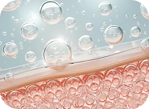
Skin aging and the appearance of signs of aging, such as the tear trough, are strongly influenced by the living environment. Among the most harmful extrinsic factors are chronic exposure to ultraviolet rays, urban pollution, tobacco, and the blue light emitted by digital screens. These often silent aggressions induce cellular oxidative stress that progressively damages collagen fibers, weakens cell membranes, and reduces elastin production.
The skin around the eyes, naturally thinner than the rest of the face, is particularly vulnerable to these attacks. It undergoes accelerated aging, resulting in dermal density loss and accentuation of hollows, such as the tear trough. Moreover, atmospheric pollution has been identified as a factor aggravating periocular pigmentation, visually enhancing the contrast between the hollow and adjacent areas.
In Geneva, where pollution levels are moderate but real, these factors must be considered in aesthetic assessment, particularly for patients exposed daily to an urban environment. Preventing these aggressions is an integral part of a comprehensive and thoughtful approach to eye-area aging.

Article written by Dr Romano Valeria
SHARE THIS ARTICLE ON